Some of these words might sound familiar, but you might not be sure of their meaning. Here are some helpful shortcuts to help your understanding of the eye and how it works.
As always, please talk to our staff if you are unsure about anything.
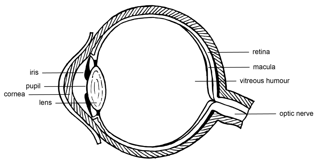
The Iris
The iris is the coloured part of the eye that controls the amount of light entering the eye by adjusting the size of the pupil.
It’s composed of muscular tissue that contracts to reduce the size of the pupil in bright light and expands to increase the size of the pupil in low light.
The unique color of each person’s iris comes from varying amounts of melanin, a pigment also responsible for skin and hair color.
The iris plays a crucial role in vision by regulating the amount of light that reaches the retina, which is where images are formed and sent to the brain.
The Pupil
The pupil is the black circular opening in the centre of the iris of the eye.
It works much like the aperture on a camera, dilating or constricting to regulate the amount of light entering the eye.
In low light conditions, the pupil expands to allow more light in and help us see better.
Conversely, in bright light, the pupil contracts to reduce the amount of light entering the eye.
The size of the pupil is controlled by the muscles in the iris.
The Cornea
The cornea is a clear membrane at the very front of your eye.
It is somewhat “pointed”, and this can be felt through the closed eyelid.
The cornea is an important structure for focussing light from the environment onto the retina.
The Lens
The lens is a clear, flexible structure located behind the iris and the pupil in the eye.
Its primary function is to focus light rays onto the retina, much like a camera lens focuses light onto a film.
It changes shape — becoming thinner or thicker — to adjust the eye’s focus for viewing objects at various distances, a process known as accommodation.
As we age, the lens can lose its flexibility, leading to common vision issues like presbyopia.
The Retina
The retina is a thin layer of tissue located at the back of the eye, directly opposite the lens.
It’s responsible for receiving light that the lens has focused, converting it into neural signals, and sending these signals to the brain for visual recognition.
The retina is composed of millions of photoreceptor cells, which are sensitive to light.
There are two types of photoreceptors: rods, which function in low light conditions, and cones, which function in bright light conditions and also perceive colour.
Dysfunction or damage to the retina can lead to significant vision impairment or blindness.
The Macula
The macula is a small, highly sensitive part of the retina responsible for detailed central vision.
It’s located in the center of the retina and contains a high concentration of cones, the light-sensitive cells that are responsible for colour perception and visual acuity.
The macula allows us to perform tasks that require sharp vision, such as reading, driving, and recognising faces.
Damage to the macula, as seen in conditions like age-related macular degeneration, can lead to loss of central vision.
The Vitreous Humour
The vitreous humour is a clear, gel-like substance that fills the space between the lens and the retina in the eye.
It makes up about two-thirds of the eye’s volume and helps maintain its round shape.
Besides providing structural support, the vitreous humour also allows light to pass through from the lens to the retina.
With age, the vitreous humour can shrink and become more liquid, sometimes leading to floaters or flashes in one’s vision.
The Optic Nerve
The optic nerve is a crucial component of the visual system.
It’s a bundle of more than a million nerve fibres that transmit visual information from the retina to the brain.
The optic nerve begins at the back of the eye, where the fibres of the retinal ganglion cells come together. This nerve carries the signals of light detected by the photoreceptor cells in the retina, enabling us to perceive images.
Damage to the optic nerve, such as in glaucoma, can lead to vision loss or blindness.
The Choroid
The choroid is a layer of the eye located between the retina and the sclera, the white part of the eye.
It’s mainly composed of blood vessels and connective tissue.
The primary function of the choroid is to provide oxygen and nutrients to the outer layers of the retina.
It also contains a pigment that absorbs excess light to prevent light reflection within the eye, thus enhancing the clarity of the visual image.
Problems in the choroid, such as inflammation or abnormal blood vessel growth, can lead to vision loss.
The Sclera
The sclera, often referred to as the ‘white of the eye,’ is the outermost layer of the eye.
It’s a tough, fibrous tissue that covers about five-sixths of the eye’s surface and extends from the cornea at the front to the optic nerve at the back.
The main functions of the sclera are to protect the eye’s internal structures and maintain its shape.
It also provides an attachment point for the muscles that move the eye.
Its white colour is due to the presence of collagen and helps to reflect excess light away from the eye.